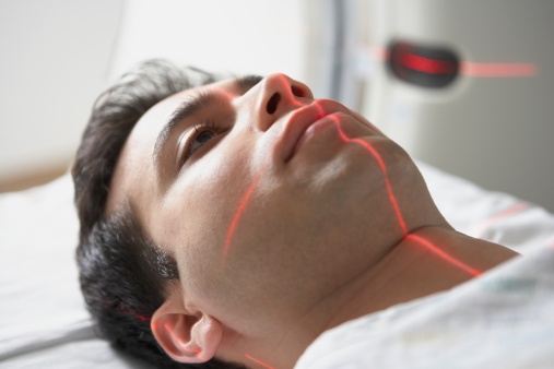

Researchers analyzed brain scans of 38 patients who had social anxiety disorders. The team discovered the scans could be used to predict, with 80% accuracy, which patients would most benefit from CBT. After the patients underwent brain scanning, they participated in 12 weeks of group-based CBT.
Researchers used “resting-state” functional magnetic resonance imaging (fMRI) to view connections to the amygdala—the part of the brain that handles fear. The team discovered that patients with a greater connectivity to the amygdala from other parts of the brain were more likely to have reduced symptoms of anxiety after CBT.
The team performed a second analysis using diffusion-weighted magnetic resonance imaging (dMRI). When they examined the dMRI scans, they found that there was a healthier connection between tracts that connect visual cues with emotional responses after receiving CBT.
Each patient was assessed before and after CBT with a behavioral assessment tool called the Liebowitz Social Anxiety Scale (LSAS). Higher LSAS scores indicated more severe social anxiety and typically associated with improvement following CBT.
Sources for Today’s Article:
Whitfield-Gabrieli, S., et al., “Brain connectomics predict response to treatment in social anxiety disorder,” Molecular Psychiatry, doi:10.1038/mp.2015.109, http://www.nature.com/mp/journal/vaop/ncurrent/full/mp2015109a.html, published online August 11, 2015.
Paddock, C., “Brain scans may help predict success of therapy for psychiatric disorders,” Medical News Today web site, August 13, 2015; http://www.medicalnewstoday.com/articles/298072.php.